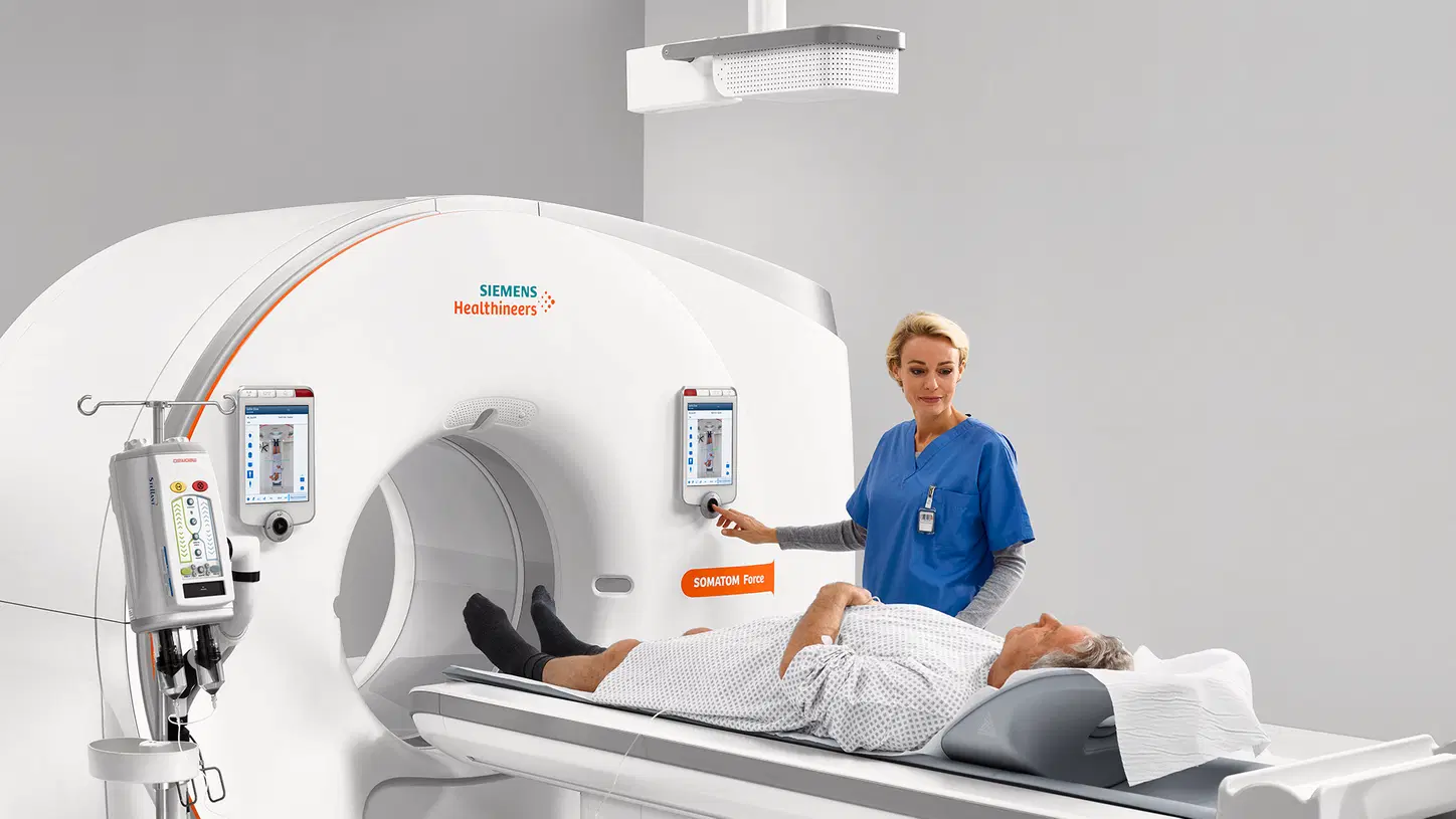
Computed tomography (CT) uses ionising radiation (X-rays). In theory, this radiation carries a very small additional risk of cancer. However, the diagnostic benefits of a CT scan performed for an appropriate medical reason are usually far greater than this small risk. The radiation dose varies depending on the body area being examined, the scanner and the imaging protocol. Avoiding unnecessary scans and keeping the dose as low as reasonably achievable (the ALARA principle) are fundamental to safe practice.
CT is a fast and painless imaging method that produces cross-sectional images of the body using X-rays taken from different angles. It provides potentially life-saving information in many situations, from medical emergencies to cancer screening. Therefore, the key question is not simply “Is CT harmful?” but rather “Is this scan necessary at this time, and has the radiation dose been optimised?”
How Is Radiation Dose Measured in CT Scans?
The effect of radiation on the body is expressed as the effective dose, measured in millisieverts (mSv). This value takes into account the varying sensitivity of different organs to radiation, allowing radiation exposure to be compared using a single measurement.
The values below are approximate averages. Actual doses may vary depending on the scanner, imaging protocol and the patient's body size.
Examination / SourceApproximate Effective Dose
Chest X-ray (PA view) | 0.1 mSv
Mammography | 0.4 mSv
Low-dose chest CT | 1–1.5 mSv
Brain CT | 2 mSv
Chest (thoracic) CT | 5–8 mSv
Abdominal and pelvic CT | 8–10 mSv
Average annual natural background radiation worldwide | 2.4–3 mSv
Istanbul–New York flight (one way) | Approximately 0.05–0.1 mSv
A practical comparison: A chest CT scan may deliver a dose roughly equivalent to 2–3 years of natural background radiation, while an abdominal CT may correspond to approximately 3–4 years. A low-dose chest CT, on the other hand, may be comparable to several months to one year of natural background radiation.
Does CT Radiation Really Cause Cancer?
Scientists estimate the risks associated with low radiation doses using the linear no-threshold (LNT) model, based on data from higher radiation exposures. According to this model, the risk is considered very small but not zero.
In general:
- A dose of 10 mSv is estimated to increase the lifetime risk of cancer by approximately 1 in 2,000.
- By comparison, the lifetime probability of developing cancer is already around 40%. A single CT scan is not expected to change this overall probability noticeably.
- These figures are estimates. It is very difficult to establish the effects of low radiation doses directly, and the model itself remains a subject of scientific discussion.
In other words, it is accurate to say that the risk is real but small. Statements such as “CT scans definitely cause cancer” or “CT scans have no harmful effects whatsoever” are both misleading.
When Does the Risk Become More Important?
Children: Developing tissues are more sensitive to radiation, and children have a longer life expectancy during which potential effects may develop. For this reason, radiation-free imaging methods such as MRI or ultrasound are generally considered first. If a CT scan is necessary, a separate low-dose protocol tailored to the child's size should be used.
Pregnancy: CT is generally avoided during pregnancy unless it is genuinely necessary for clinical reasons. Ultrasound and MRI are the preferred options where appropriate. However, if a CT scan is required in a life-threatening situation, such as trauma or suspected pulmonary embolism, the decision is made by carefully weighing the potential benefits and risks. If you are pregnant or think you may be pregnant, be sure to inform the medical team before the scan.
Repeated scans within a short period: Cumulative radiation exposure should be taken into account. Showing your previous imaging examinations and reports to your radiologist can help prevent unnecessary repeat scans.
What Measures Help Reduce Radiation Dose?
Safe CT practice is based on three key principles:
1. Justification: Is the scan genuinely necessary? Could the same diagnostic information be obtained using a radiation-free method such as MRI or ultrasound?
2. Optimisation: Imaging protocols should be adjusted according to the patient's age, weight and the body area being examined. Modern CT scanners use techniques such as automatic dose modulation, iterative image reconstruction and examination-specific scan ranges to reduce radiation exposure while maintaining image quality.
3. Dose monitoring: Radiation doses should be recorded, monitored and reviewed through regular quality-control procedures.
When Should You Ask Questions About a CT Scan?
You should feel comfortable asking questions in the following situations:
- If your doctor has not explained why the scan is necessary, do not hesitate to ask. A good physician should be able to explain the medical reason.
- If you have recently undergone a CT scan of the same body area at another centre, bring your previous images and report.
- If you are pregnant or think you may be pregnant, inform your medical team.
- If a CT scan has been recommended for your child, ask whether MRI or ultrasound could be suitable alternatives.
Is Avoiding CT Scans Always the Safer Option?
No. Avoiding a medically necessary CT scan may delay the diagnosis of serious conditions such as cancer, stroke, internal bleeding, pulmonary embolism or appendicitis. In these situations, the risks associated with delayed diagnosis may be far greater than the theoretical risk from radiation.
What matters is that the scan is performed for the right medical reason, using an appropriate protocol and at the right time.
Common Misconceptions About Radiation
“Radiation remains in the body after a CT scan.”
No. Radiation is delivered during the scan itself. No radioactive material remains in your body afterwards, and you do not emit radiation to people around you.
“MRI and CT are the same; both use radiation.”
No. MRI uses magnetic fields and radio waves. It does not involve ionising radiation.
“Contrast material means radiation.”
No. The iodine-based contrast agent used in CT scans does not increase the radiation dose. It helps certain tissues and structures appear more clearly in the images.
“Having one CT scan every year is always safe.”
A single scan generally involves a relatively low radiation dose. However, unnecessary repeat examinations and cumulative exposure should be avoided. The most important consideration is whether each scan is medically justified.
Frequently Asked Questions About CT Scans
How many X-rays is one CT scan equivalent to?
A brain CT scan may deliver a radiation dose equivalent to approximately 20 chest X-rays. A chest CT may correspond to around 50–80 chest X-rays, while an abdominal CT may be equivalent to approximately 80–100 chest X-rays. These figures are estimates and vary depending on the scanner and imaging protocol.
Do I need to take any precautions after a CT scan?
No special precautions are necessary in terms of radiation exposure. If you have had a contrast-enhanced CT scan, drinking plenty of water is generally recommended, unless your doctor has advised otherwise.
Can I have a CT scan while breastfeeding?
The X-rays used in CT scans do not affect breast milk production or the safety of breast milk. When iodine-based contrast material is used, only a very small amount passes into breast milk. Current guidelines generally do not recommend interrupting breastfeeding after the use of standard iodinated contrast agents. Nevertheless, consult your radiologist and doctor before the examination.
Who is eligible for low-dose chest CT screening?
Low-dose chest CT is generally considered for lung cancer screening in people within certain age groups who have a significant smoking history. Your doctor will determine whether you meet the eligibility criteria.
Can I find out the radiation dose before my CT scan?
Imaging centres record the radiation dose delivered by their scanners. If you would like to know the dose, you can ask your radiologist before or after the examination.
Conclusion
Computed tomography uses ionising radiation and carries a small potential risk. However, when performed for an appropriate medical reason, with patient-specific dose optimisation and under the supervision of experienced radiologists, its diagnostic benefits generally far outweigh this risk.
Rather than postponing or refusing a scan, the most sensible approach is to ask why it is needed and share details of any previous imaging examinations with your doctor.
At Centermed, CT examinations are performed under the supervision of experienced radiologists, with careful consideration of medical necessity and patient safety.
For information and appointments, call 444 2 211 or contact us via WhatsApp at +90 530 413 96 48.
This article is intended for general informational purposes only and does not replace medical advice. Please consult your doctor to determine whether a CT scan is necessary and to assess your individual risk. The radiation dose figures provided are approximate averages.