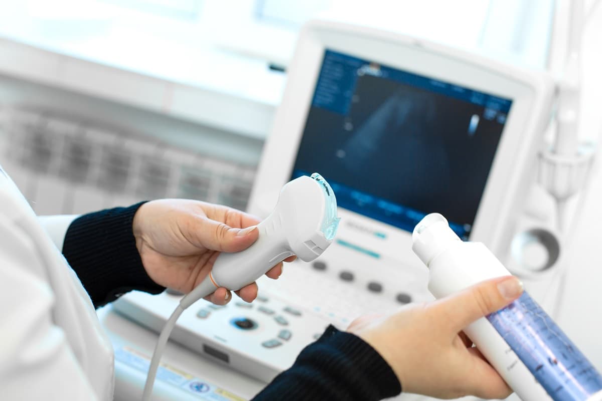
Ultrasound (USG) is commonly used as a first-line imaging method for a wide range of symptoms and conditions, including abdominal pain, bloating, kidney and gallbladder problems, swelling in the neck or thyroid abnormalities, palpable breast lumps or breast pain, and pain involving the joints, tendons and muscles.
Ultrasound does not use ionising radiation. It is painless, quick and provides real-time images. For these reasons, it can also be safely used in pregnant women and children. However, ultrasound is not sufficient for every condition. In some cases, further evaluation with MRI, computed tomography (CT) or mammography may be necessary.
Ultrasound imaging uses sound waves that bounce off body tissues to create images. Doctors often prefer ultrasound as an initial examination because it is fast, accessible and safe. In this guide, we explain the four most common areas of ultrasound use and the symptoms that may indicate a need for each examination.
Why Is Ultrasound So Widely Used?
- It does not use ionising radiation. The examination can be repeated and is suitable for use during pregnancy and childhood.
- It provides real-time images. Organ movement, blood flow (Doppler ultrasound) and fluid accumulation can be observed as they happen.
- It guides medical procedures. Ultrasound can help guide needle biopsies, fluid drainage and other minimally invasive procedures.
- It is quick and accessible. Most examinations take 10–30 minutes and generally do not require special preparation.
Limitations: Structures located behind bones or air-filled organs cannot be visualised clearly. Image quality may also be reduced in patients with obesity or excessive intestinal gas. In such cases, another imaging method may be recommended.
1. When Is an Abdominal Ultrasound Recommended?
Abdominal ultrasound is used to examine the liver, gallbladder and bile ducts, pancreas, spleen, kidneys, urinary bladder and major blood vessels within the abdomen.
Common reasons for requesting an abdominal ultrasound include:
- Pain in the upper right abdomen, particularly pain that worsens after eating fatty foods (possible gallstones).
- Abdominal bloating or a persistent feeling of fullness.
- Flank or lower back pain resembling renal colic, or blood in the urine.
- Jaundice or elevated liver enzyme levels.
- Assessment of fatty liver disease.
- Frequent urination, difficulty passing urine, or suspected kidney or bladder stones.
- A palpable lump or swelling in the abdomen.
- Chronic abdominal pain or unexplained weight loss.
Preparation: For examinations of the upper abdomen, fasting for 6–8 hours is generally required. For bladder and pelvic examinations, a full bladder may be necessary. Preparation instructions may vary between medical centres, so please confirm the requirements when booking your appointment.
Note: In cases of suspected appendicitis, serious trauma or severe abdominal pain, ultrasound may be used as the initial examination. However, if the findings are inconclusive, CT or MRI may be required.
2. When Is a Thyroid Ultrasound Recommended?
A thyroid ultrasound examines the size and structure of the thyroid gland, located at the front of the neck, and helps identify nodules or other abnormalities within it.
Common reasons for requesting a thyroid ultrasound include:
- A palpable swelling in the neck or a lump noticed when swallowing.
- Difficulty swallowing, a sensation of something stuck in the throat, or hoarseness.
- A diagnosis of hyperthyroidism or hypothyroidism, or abnormal thyroid-stimulating hormone (TSH) levels.
- A family history of thyroid cancer or thyroid disease.
- Monitoring a previously detected thyroid nodule.
- An incidental thyroid nodule identified during another examination, such as CT, MRI or PET.
What does the ultrasound show?
The examination assesses the size, margins and structure of a nodule (whether it is cystic or solid), as well as features such as calcifications and blood flow. These findings help determine whether a biopsy is necessary.
Most thyroid nodules are benign. The decision to perform a biopsy is based on the ultrasound findings and the size of the nodule. When necessary, fine-needle aspiration biopsy (FNAB) can be performed under ultrasound guidance.
Preparation: No special preparation is usually required.
3. When Is a Breast Ultrasound Recommended?
Breast ultrasound is an important complementary imaging method, particularly for younger women and women with dense breast tissue. It is often used alongside mammography to provide a more comprehensive assessment.
Common reasons for requesting a breast ultrasound include:
- A palpable lump or area of thickening in the breast.
- Breast pain or persistent tenderness in a specific area.
- Nipple discharge, particularly bloody or one-sided discharge.
- Skin dimpling, redness or changes in breast shape.
- Swelling in the armpit or enlarged lymph nodes.
- Further evaluation of a finding detected on mammography.
- Initial imaging for breast-related symptoms in women under 40.
- Additional assessment alongside mammography in women with dense breast tissue.
- Evaluation of breast implants.
Mammography or ultrasound?
These two methods are not alternatives to each other; they complement one another. Mammography is particularly effective at detecting microcalcifications, while ultrasound is useful for distinguishing fluid-filled cysts from solid masses. In certain cases, breast MRI may also be recommended.
You can read more about this examination in our article, “What Is Breast MRI?”
Timing: In women who menstruate, breast tissue is generally less sensitive during the first half of the menstrual cycle. However, if a new lump is detected, medical evaluation should not be delayed until a particular point in the cycle.
4. When Is a Musculoskeletal Ultrasound Recommended?
Musculoskeletal ultrasound provides high-resolution images of muscles, tendons, ligaments, joint capsules, nerves and fluid within the joints. It also allows dynamic assessment of joints and soft tissues, meaning they can be examined while moving.
Common reasons for requesting a musculoskeletal ultrasound include:
- Shoulder pain and difficulty raising the arm (rotator cuff injuries, bursitis and tendinitis).
- Elbow pain (tennis elbow or golfer's elbow).
- Numbness in the wrist and fingers, or suspected carpal tunnel syndrome.
- Heel pain (plantar fasciitis) or Achilles tendon problems.
- Swelling and fluid accumulation around the knee, hip or ankle.
- Muscle tears and tendon or ligament injuries in athletes.
- Palpable soft-tissue masses, such as cysts, lipomas and ganglion cysts.
- Screening for developmental dysplasia of the hip in infants.
Why might an MRI also be needed to assess internal joint structures?
MRI is more effective for evaluating deeper structures, such as the menisci and cruciate ligaments inside the knee, as well as bone marrow oedema and joint cartilage.
Ultrasound is particularly effective for examining superficial tendons, ligaments, muscles and fluid collections. The most appropriate imaging method is determined by considering the clinical findings together with the radiologist's assessment.
Image-guided procedures: Joint injections, injections around tendons and fluid aspiration can be performed under ultrasound guidance.
Which Imaging Method Is Suitable for Which Symptom? (Summary Table)
Symptom or suspected conditionFirst-line examinationAdditional examination if needed
Pain in the upper right abdomen | Abdominal ultrasound | CT, MRI / MRCP
Suspected kidney stones | Abdominal ultrasound | Low-dose CT
Neck swelling or thyroid nodule | Thyroid ultrasound | FNAB, thyroid scintigraphy
Palpable breast lump (under 40) | Breast ultrasound | MRI, biopsy
Palpable breast lump (over 40) | Mammography + breast ultrasound | MRI, biopsy
Shoulder pain | Shoulder ultrasound | Shoulder MRI
Suspected meniscus or ligament injury in the knee | X-ray, MRI | Ultrasound (for swelling or fluid)
Abdominal pain during pregnancy | Ultrasound | MRI
This table is intended as a general guide. The final decision is always made by the physician based on the patient's clinical assessment.
General Preparation Before an Ultrasound Examination
- Abdominal ultrasound: Fasting for 6–8 hours; a full bladder may sometimes also be required.
- Pelvic/lower abdominal ultrasound: A full bladder is usually required. This can generally be achieved by drinking 3–4 glasses of water approximately one hour before the examination.
- Thyroid, breast and musculoskeletal ultrasound: No special preparation is usually required.
- Bring your previous examination results and medical reports, as they are valuable for comparison.
- Wear comfortable clothing that can be easily removed or adjusted.
What Happens After an Ultrasound Examination?
Most examinations take 10–30 minutes, and you can return to your daily activities immediately afterwards.
The radiologist reviews the images and prepares a report. You should discuss the findings with your treating physician, who can interpret them in the context of your symptoms and medical history. If necessary, additional imaging or a biopsy may be recommended.
Frequently Asked Questions
Is ultrasound harmful?
No. Ultrasound does not use ionising radiation and has no known harmful effects when used for medical purposes. It can also be safely performed during pregnancy.
Can cancer be diagnosed with ultrasound?
Ultrasound provides important information about the structure and characteristics of certain masses. However, a definitive diagnosis is often made through biopsy and pathological examination. If suspicious findings are detected, further investigations may be recommended.
Do I need to fast before an abdominal ultrasound?
In most cases, yes, particularly when the upper abdominal organs are being examined. Fasting helps keep the gallbladder full and reduces intestinal gas. Be sure to confirm the preparation instructions when booking your appointment.
If I have breast pain, should I have an ultrasound or a mammogram?
The choice depends on your age and clinical findings. Ultrasound is generally used for women under 40, while mammography and ultrasound are often used together for women over 40. Your doctor will recommend the most appropriate examination.
My ultrasound results are normal, but my symptoms persist. What should I do?
A normal ultrasound result does not rule out every possible condition. If your symptoms continue, consult your doctor. Other imaging methods, such as MRI or CT, may be necessary.
When will my ultrasound results be available?
At most medical centres, results are available shortly after the examination. Centermed patients can also access their results through the online results system.
Conclusion
Ultrasound is a safe, quick and informative first step in evaluating conditions involving the abdomen, thyroid gland, breasts and musculoskeletal system. However, ultrasound does not provide the answer to every medical concern. The most appropriate imaging method depends on your symptoms, age and clinical condition.
If you notice any symptoms or findings that cause concern, do not delay seeking medical advice.
At Centermed, ultrasound examinations are performed by experienced radiologists. When necessary, the findings are evaluated alongside MRI, CT or mammography results to provide a more comprehensive assessment.
For information and appointments, please call:
444 2 211
Or contact us via WhatsApp:
+90 530 413 96 48
This article is intended for general informational purposes only and does not replace medical advice. Please consult your doctor for diagnosis and treatment.