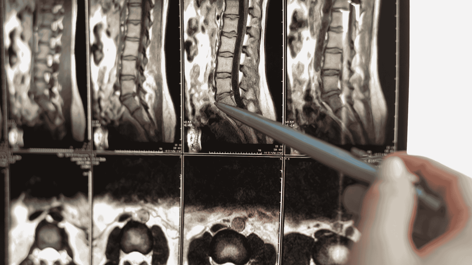
Lumbar CT (Computed Tomography) is a medical imaging method used to visualize the lumbar spine (lower back) in detail. This procedure is typically performed using a CT scanner and allows for the examination of the bones, discs, spinal canal, and surrounding tissues in the lumbar region.
The lumbar region is the lower part of the spine and consists of five vertebrae (L1–L5). It is the main part of the spine responsible for bearing body weight and is often subjected to stress in daily life, making it a common area for pain and discomfort. Lumbar CT is especially used to identify the source of complaints such as lower back pain, sciatic pain (pain radiating to the legs), movement limitations, or post-traumatic conditions.
When is Lumbar CT Requested?
- Suspected herniated disc
- Spinal fractures
- Degenerative disc disease
- Spondylolisthesis (slipped vertebra)
- Spinal tumors
- Spinal canal stenosis
- Infections (discitis, osteomyelitis)
- Congenital spinal anomalies
- Post-surgical follow-up
How is it Performed?
During a lumbar CT scan, the patient lies on their back and is moved into the scanner. The CT machine uses X-rays to capture multiple cross-sectional images of the lower back. These images are combined by a computer to form 3D structures. The procedure usually takes 5–10 minutes and is painless.
In some cases, a contrast agent (dye) may be used to enhance image clarity or distinguish certain tissues. This dye is administered intravenously and is generally safe, though precautions may be needed for individuals with allergies.
What is the Difference Between Lumbar CT and MRI?
- CT (Computed Tomography) is more effective for assessing bone structures and is thus preferred for detecting fractures and deformities.
- MRI (Magnetic Resonance Imaging) provides more detailed images of soft tissues (discs, nerves, ligaments).
Depending on the suspected condition, a physician may choose CT or MRI, or sometimes both may be requested.
Conclusion
Lumbar CT is a highly effective imaging method for diagnosing structural problems in the lower back. It is an important tool for identifying the cause of pain, trauma, or suspected spinal diseases, and for planning appropriate treatment. Since it involves radiation, it is not recommended for pregnant women or unnecessary cases. However, when used appropriately, it provides fast and accurate results.